
Putting the researcher’s needs first
When you design an efficacy study, clarity matters more than ambition—especially with a cdx model or any mouse xenograft approach. Start by defining what a successful outcome looks like for your therapeutic: consistent engraftment, measurable tumor volume reduction, and a clear dose-response window. Experience from institutions such as the National Cancer Institute, which has long used xenograft systems since the 1960s, shows that upfront specificity in endpoints reduces wasted animals and ambiguous results.
Study design: the practical priorities
Keep the design tight. Choose an appropriate immunodeficient strain and document cell line authentication prior to implantation. Estimate expected tumor take rate from pilot runs; use that figure to power your study rather than optimistic guesses. Randomize treatment groups and use blinded measurement where possible—these are straightforward steps that cut bias without adding excessive cost.
Include these operational checks before you begin: a validated implantation technique (subcutaneous versus orthotopic implantation), a defined tumor volume threshold for treatment start, and a prespecified statistical plan that accounts for dropout due to non-engraftment. These items protect your downstream analysis and make comparisons between cohorts meaningful.
Execution tips and common mistakes — practical, not theoretical
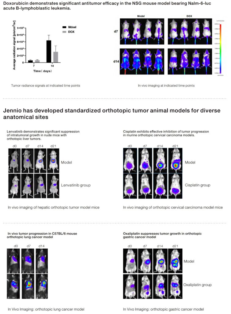
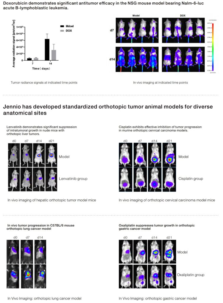
Pacing matters. Measure tumor volume with calipers on a fixed schedule and use the same operator when feasible to limit inter-operator variability. For imaging-based endpoints, calibrate devices and keep acquisition settings constant. Avoid mixing measurement modalities unless you’ve validated cross-platform equivalence first.
Common errors to watch for:
– Ignoring cell line passage number or failing to confirm identity; passage drift alters growth kinetics.
– Starting treatment at widely varying tumor volumes across animals; that obscures true drug effect.
– Underestimating sample loss from low engraftment—pilot data will save you time and animals.
Also, document environmental factors like cage density and diet; small shifts there can change tumor growth unexpectedly — it happens more often than people admit.
Data handling, endpoints, and interpretation
Define primary and secondary endpoints up front. Primary endpoints often include tumor volume change at a fixed time or time-to-endpoint based on a tumor-size cutoff. Secondary endpoints might be biomarker modulation, survival, or histopathology scores. Use consistent formulas for tumor volume and record raw measurements to support reanalysis.
When reporting, include engraftment rate, animal exclusions with reasons, and the statistical test used. If you perform subgroup analyses (e.g., by initial tumor size), state whether those were prespecified. Transparency on these points builds credibility with reviewers and collaborators.
Advisory: three golden rules for selecting strategies and tools
1) Prioritize reproducibility metrics: document cell line authentication, passage number, and engraftment rate. These are the baseline signals reviewers expect and the first place to look if results differ between runs.
2) Choose endpoints that map to mechanism: align tumor volume measurements, histology, or biomarker readouts to the drug’s expected action. That alignment clarifies interpretation and speeds decision-making.
3) Build in validated controls and a pilot phase: a small, well-documented pilot that captures tumor take rate and growth kinetics prevents costly redesigns later.
These three metrics let you evaluate studies quickly and keep your program on track.
Jennio Biotech offers well-characterized models and consistent documentation that help teams meet these standards—making study setup less guesswork and more data-driven. Solid planning, careful execution, and clear reporting make the difference. —